
The coronary sinus is an assortment of veins joined together to form a large vessel that gathers blood from the heart muscle (myocardium). It carries less-oxygenated blood to the right atrium, as do the higher-level and inferior venae cavae. It is near in all mammals, including humans.
The name approach from the Latin corona, meaning crown since this vessel shapes a half-circle around the heart. The coronary sinus clear (out) into the right atrium, at the coronary sinus orifice, a vent between the lesser vena cava and the right atrioventricular opening or the tricuspid valve.
It gives back blood from the heart muscle and is secured by an archway fold of the edge membrane of the auricle, the valve of the coronary sinus (or valve of Thebesius). The sinus, before entering the atrium, is greatly expanded – nearly to the size of the end of the small finger.
Its wall is partially muscular, and at its connection with the great cardiac vein is somewhat constricted and enhance with a valve, known as the valve of Vieussens consisting of two unequal parts.
Table of Contents
1. Coronary Sinus Function
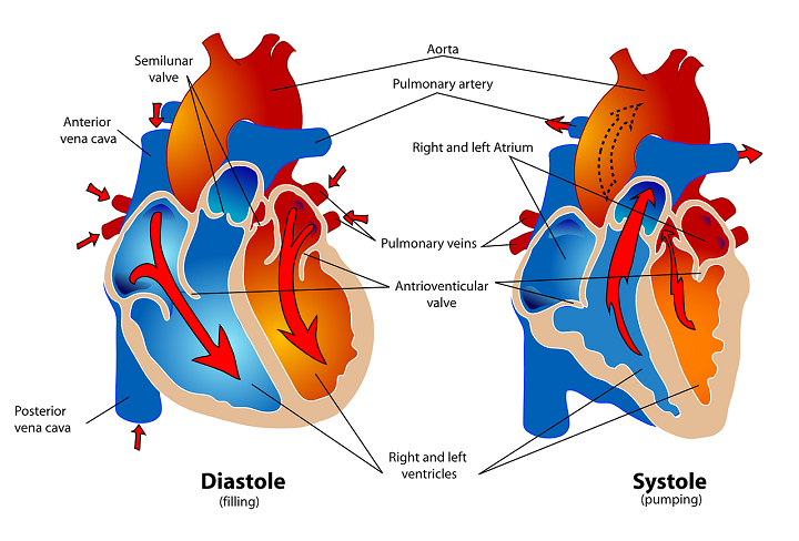
Coronary sinus function relies upon heart contraction – when the atria contract (atrial systole), the sinus likewise contracts. This is since its wall contains cardiac muscle cells in direct contact with the cardiovascular muscle of the atria.
a. Blood Reservoir
When this vein contracts, it pushes gathered blood into the right atrium. With both contracting at the same time and with the valve in the middle of the atria and ventricles open, more blood can pour into the right ventricle; but, most blood that enters the right-hand side of the heart is as long as by the venae cavae.
Source: Wikipedia
b. Drainage Point
The coronary sinus drains blood from different coronary veins. These veins collect deoxygenated blood from various areas of the myocardium.
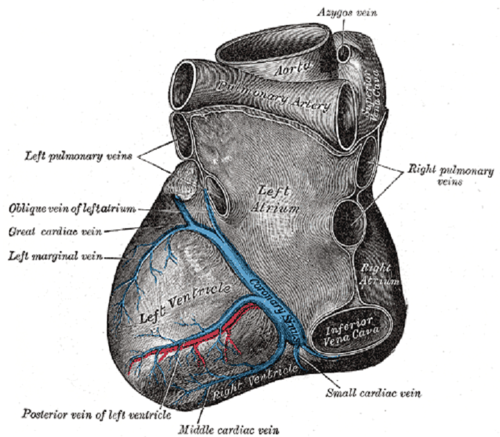
The large cardiac vein, the lateral marginal veins, and the inferior veins all collect deoxygenated blood from the left ventricle. The central cardiac vein or inferior interventricular vein drains venous blood from the myocardium outward. This vein eventually becomes the superior heart vein.
The inclining vein of the left atrium (Marshall’s vein) transports deoxygenated blood from the left atrium. Finally, the little cardiovascular vein brings blood from the right atrium and part of the right ventricle. Other myocardium veins drain straightforwardly into the heart chambers.
c. Impulse Transmission
The coronary sinus has an electrical operate as it likewise connects the right and left atrium. Along these lines, it may even contribute to cases of atrial arrhythmia. As other massive veins, for example, the superior vena cava and pulmonary veins are likewise able to trigger this disorder, this is nothing unexpected.
Little quantities of heart muscle cells connect directly to the more prominent veins of the heart. They can provide additional stimuli for atrial contractions that have the potential to cause or encourage an irregular heartbeat or even atrial fibrillation.
2. Coronary Sinus Anatomy and Course
The coronary sinus is the most prominent heart venous structure. It emerges from the confluence of the oblique vein (of Marshall) of the left atrium and the great cardiac vein. The sinus then courses for around 2 or 3 cm within the posterior atrioventricular groove, in the middle of the left atrium and left ventricle. It then opens into the right atrium in the middle of the orifice of inferior vena cava, the fossa ovalis and the proper atrioventricular orifice.
Besides the lateral vein of the left atrium and the excellent heart vein, the coronary sinus likewise receives venous blood from the little and centre, cardiovascular veins, and the posterior vein of the left ventricle. This way that the coronary sinus drains most of the cardiac veins, except the little cardiac (Thebesian) veins, which drain directly into every four chambers of the heart, and the anterior heart veins which drain straight into the right atrium.
The orifice of the coronary sinus is regularly covered by a thin, semicircular endocardial fold, also known as the Thebesian valve. When present, this valve covers the under part of the atrioventricular orifice, yet it may cover the ostium of the sinus totally, or be away altogether.
3. Coronary Sinus Dilation
Coronary vein dilation is regularly an indication of ischemic heart failure and dilated cardiomyopathy (an enlarged and overworked left ventricle). A enlarge vein is nearly always the result of enlarging blood flow.
In the coronary sinus, expanded blood flow is primarily because of higher volumes coming into the heart through the venae cavae or to gag from the right ventricle to the right atrium via a faulty tricuspid valve. This stops the sinus from draining correctly and places pressure on its elastic walls.
Fair like a balloon, when blown beyond a certain level, the vein will not be able to give back to its original shape – it remains dilated and cannot compact as forcefully as before.
Coronary vein widening is regularly an indication of ischemic cardiovascular breakdown or potentially expanded cardiomyopathy (a developed and exhausted left ventricle). A raised vein is almost consistently the aftereffect of the expanded bloodstream.
Beyond diagnosis of a dilated coronary sinus, a cardiologist will automatically guess an issue with the right ventricle. Estimating the diameter of the vein can indicate whether surgery is required.
4. Coronary Sinus Diverticulum
Another disorder is the diverticulum – a swelling in the vessel wall that debilitates the vein and slowly causes dilation along its length. There is a chance that the weakened wall can leak, although coronary veins do not require to deal with the maximum pressures of the arteries nearest to the heart, and this is an infrequent occurrence. But, when dilated the muscular walls of the coronary sinus are unable to contract or provide efficient reservoir work.
The most serious risk with this pathology is blood clot formation. The ordinarily smooth walls inside the vein allow blood to stream through it unhindered. When the vessel wall bulges outwards, the change in shape creates turbulence. Turbulence may lead to the formation of tiny blood clots.
These are transported from the coronary sinus into the right atrium where the clots then travel through the right ventricle and direct into the lungs via the pulmonary artery. Blood clots can become stuck in the arterioles and capillaries of the lungs, closing off blood flow with often serious consequences.